Tricuspid: Difference between revisions
No edit summary |
No edit summary |
||
| (12 intermediate revisions by 2 users not shown) | |||
| Line 1: | Line 1: | ||
==Anatomy== | ==Anatomy== | ||
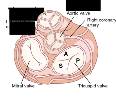
The tricuspid valve, ensures that there is no backflow of blood from the right ventricle to the right atrium. The tricuspid valve is like the Latin name indicates consists of three leaflets: the septal (S), the anterior (A) and posterior (P) leaflet, whose anterior leaflet is the largest. The tricuspid valve, distinguishes itself not only by the number of valve leaflets of the mitral valve, but also by the manner of attachment. The chordae of the mitral valve attached to two papillary cups which the tricuspid valve attached to a lot more muscular and cups directly to the septum. The valve is located slightly to the apex than the mitral valve. | The tricuspid valve, ensures that there is no backflow of blood from the right ventricle to the right atrium. The tricuspid valve is like the Latin name indicates consists of three leaflets: the septal (S), the anterior (A) and posterior (P) leaflet, whose anterior leaflet is the largest. The tricuspid valve, distinguishes itself not only by the number of valve leaflets of the mitral valve, but also by the manner of attachment. The chordae of the mitral valve attached to two papillary cups which the tricuspid valve attached to a lot more muscular and cups directly to the septum. The valve is located slightly to the apex than the mitral valve.<cite>1</cite> | ||
[[Image: | [[Image:Tvalve.svg|400px]] | ||
==Echocardiographic views== | ==Echocardiographic views== | ||
| Line 14: | Line 14: | ||
|- | |- | ||
|[[Image:Ap4ch.jpg|400px]] | |[[Image:Ap4ch.jpg|400px]] | ||
|[[Image: | |[[Image:Subcostaal4ch03.jpg|400px]] | ||
|- | |- | ||
!AP4CH | !AP4CH | ||
| Line 20: | Line 20: | ||
|} | |} | ||
==Stenosis== | |||
An isolated tricuspid valve stenosis is extremely rare in adulthood. Almost always there is a combination with more or less serious tricuspid valve insufficiency. When a mitral valve stenosis is present in old age the cause is almost always a previously created rheumatic fever. In 10% of the patients, also, there appears to be a tricuspid valve stenosis. Dooming the valve leaflets is always an expression of a tricuspid valve stenosis. For assessing the severity of a tricuspid valve stenosis no objective criteria exists, however it is important to realize that the pressure differences in the tricuspid valve stenosis will be less. At a mean gradient of 5 or greater may be a serious tricuspid valve stenosis. The peak gradient is high, both in patients with a severe stenosis without TI as in patients with severe TS with low TI. The deceleration time will show obvious differences. | |||
Click [[Tricuspid Stenosis|'''here''']] for quantification tricuspid valve stenosis. | |||
==Insufficiency== | |||
Tricuspid insufficiency (TI) is most common on the basis of RA or RV dilatation. Also, there may occur failure in inferior infarction (proximal RCA stenosis). A tricuspid insufficiency rarely is caused by damage to the valve itself. Typically, there is a functional TI, which is the result of the RV and dilatation of the annulus. | |||
{| class="wikitable" cellpadding="0" cellspacing="0" border="0" width="500px" | |||
|- | |||
|colspan="2"|'''Causes TI''' | |||
|- | |||
!Functional TI | |||
| | |||
*disorders of the right ventricle: RV infarction, dilated cardiomyopathy | |||
*secondary to pulmonary hypertension, cor pulmonale, for example, a pulmonary embolism or primary. | |||
*mitral stenosis or mitral valvular insufficiency | |||
*left-right bypass, such as an atrial septal defect or ventricular septal defect a | |||
*Eisenmenger syndrome (rare) | |||
*pulmonary stenosis | |||
*hyperthyroidism | |||
|- | |||
!Secondary TI | |||
| | |||
*Ebstein anomaly | |||
*infective endocarditis | |||
*Trauma . | |||
*rheumatic fever | |||
*carcinoid | |||
*Papillary muscle disorders | |||
*Connective tissue diseases such as Marfan Syndrome. | |||
*Non-infectious endocarditis, such as in patients with SLE or rheumatoid arthritis | |||
*Damage caused by the electrode of a pacemaker or ICD | |||
|} | |||
The symptoms of TI are due to the decompensated right heart failure: peripheral edema, ascites, pleural effusion, an enlarged liver and / or spleen, increased CVD and often poor appetite, sometimes with cachexia and malnutrition as a result. In addition, there is generally manifestations of forward failure: exercise-induced shortness of breath, fatigue , cold fingers or toes . | |||
As tricuspid insufficiency almost always is the result of an underlying disease, treatment is aimed at eliminating the underlying cause. Sometimes a repair of tricuspid valve is provided, this usually consists of a plastic tricuspid valve. | |||
Click [[Tricuspid Insufficiency|'''here''']] for quantification tricuspid insufficiency . | |||
==References== | ==References== | ||
<biblio> | <biblio> | ||
#1 | #1 [http://www.geisinger.kramesonline.com/3,S,89116 Geisinger - How the Heart Works] | ||
</biblio> | </biblio> | ||
Latest revision as of 20:28, 7 February 2014
Anatomy
The tricuspid valve, ensures that there is no backflow of blood from the right ventricle to the right atrium. The tricuspid valve is like the Latin name indicates consists of three leaflets: the septal (S), the anterior (A) and posterior (P) leaflet, whose anterior leaflet is the largest. The tricuspid valve, distinguishes itself not only by the number of valve leaflets of the mitral valve, but also by the manner of attachment. The chordae of the mitral valve attached to two papillary cups which the tricuspid valve attached to a lot more muscular and cups directly to the septum. The valve is located slightly to the apex than the mitral valve.[1]
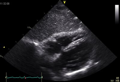
Echocardiographic views
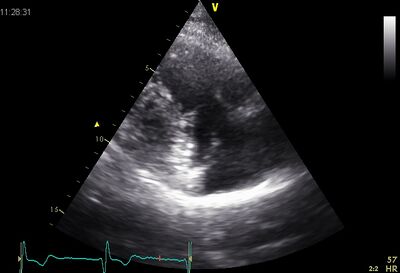
|
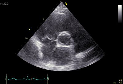
|
| Plax by tilted | PSax Ao |
|---|---|
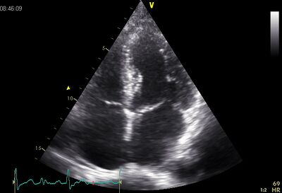
|
|
| AP4CH | Subcostal |
Stenosis
An isolated tricuspid valve stenosis is extremely rare in adulthood. Almost always there is a combination with more or less serious tricuspid valve insufficiency. When a mitral valve stenosis is present in old age the cause is almost always a previously created rheumatic fever. In 10% of the patients, also, there appears to be a tricuspid valve stenosis. Dooming the valve leaflets is always an expression of a tricuspid valve stenosis. For assessing the severity of a tricuspid valve stenosis no objective criteria exists, however it is important to realize that the pressure differences in the tricuspid valve stenosis will be less. At a mean gradient of 5 or greater may be a serious tricuspid valve stenosis. The peak gradient is high, both in patients with a severe stenosis without TI as in patients with severe TS with low TI. The deceleration time will show obvious differences.
Click here for quantification tricuspid valve stenosis.
Insufficiency
Tricuspid insufficiency (TI) is most common on the basis of RA or RV dilatation. Also, there may occur failure in inferior infarction (proximal RCA stenosis). A tricuspid insufficiency rarely is caused by damage to the valve itself. Typically, there is a functional TI, which is the result of the RV and dilatation of the annulus.
| Causes TI | |
| Functional TI |
|
|---|---|
| Secondary TI |
|
The symptoms of TI are due to the decompensated right heart failure: peripheral edema, ascites, pleural effusion, an enlarged liver and / or spleen, increased CVD and often poor appetite, sometimes with cachexia and malnutrition as a result. In addition, there is generally manifestations of forward failure: exercise-induced shortness of breath, fatigue , cold fingers or toes .
As tricuspid insufficiency almost always is the result of an underlying disease, treatment is aimed at eliminating the underlying cause. Sometimes a repair of tricuspid valve is provided, this usually consists of a plastic tricuspid valve.
Click here for quantification tricuspid insufficiency .
