Parasternal long axis: Difference between revisions
Jump to navigation
Jump to search
No edit summary |
No edit summary |
||
| Line 12: | Line 12: | ||
==Schematic view== | ==Schematic view== | ||
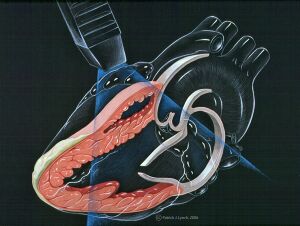
Position of the transducer on the chest and schematic echocardiographic view | Position of the transducer on the chest and schematic echocardiographic view | ||
{{clr}} | |||
==Example== | ==Example== | ||
This a parasternal long axis view of a normal heart. | This a parasternal long axis view of a normal heart. | ||
Revision as of 11:07, 21 September 2007
Content is incomplete and may be incorrect. |
| Author | I.A.C. van der Bilt | |
| Moderator | I.A.C. van der Bilt | |
| Supervisor | ||
| some notes about authorship | ||
The left parasternal imaging planes are found by placing the transducer in the third or fourth intercostal space on the left of the sternum.
The left parasternal long axis is usually the first view of the echocardiographic examination.
Schematic view
Position of the transducer on the chest and schematic echocardiographic view
Example
This a parasternal long axis view of a normal heart.
| <flash>file=PSLAX.swf |
| A parasternal long axis |
