Pulmonary Artery: Difference between revisions
Jump to navigation
Jump to search
No edit summary |
No edit summary |
||
| Line 7: | Line 7: | ||
|- | |- | ||
|bgcolor="#FFFFFF"|[[Image:Pulmart.png|200px]] | |bgcolor="#FFFFFF"|[[Image:Pulmart.png|200px]] | ||
|} | |||
{| class="wikitable" cellpadding="0" cellspacing="0" border="0" width="600px" | |||
|- | |||
|colspan="5"|'''PA diameter''' | |||
|- | |||
| | |||
!Reference range | |||
!Mildly abnormal | |||
!Moderately abnormal | |||
!Severely abnormal | |||
|- | |||
!1cm Below pulmonic valve | |||
|1.5–2.1 | |||
|2.2–2.5 | |||
|2.6–2.9 | |||
|≥3.0 | |||
|} | |||
{| class="wikitable" cellpadding="0" cellspacing="0" border="0" | |||
|- | |||
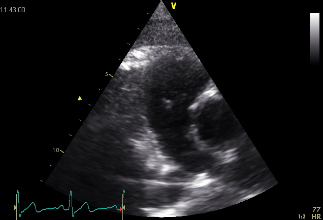
|[[Image:Pulm art03.jpg]] | |||
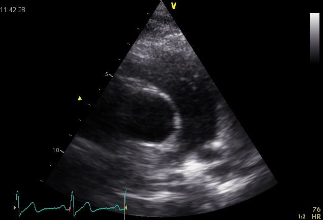
|[[Image:Pulm art02.jpg]] | |||
|- | |||
|Plax by tilted (Plax PV) | |||
|PSax Ao | |||
|- | |||
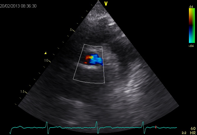
|[[Image:Pulmart03.jpg]] | |||
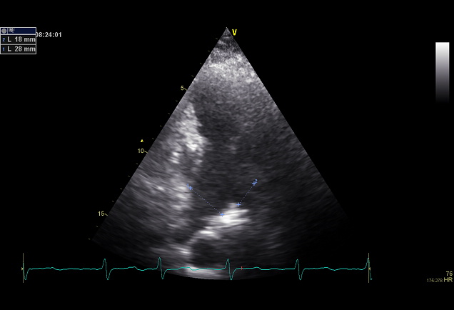
|[[Image:Pulmart02.jpg]] | |||
|- | |||
|Suprasternal apd (color doppler) | |||
|Dilated apd (Plax PV) | |||
|} | |} | ||
| Line 12: | Line 44: | ||
<biblio> | <biblio> | ||
#1 pmid=20233780 | #1 pmid=20233780 | ||
#2 pmid= | #2 pmid=3730205 | ||
</biblio> | </biblio> | ||
Revision as of 07:38, 7 January 2014
Anatomy
The pulmonary artery carries deoxygenated blood from the right ventricle to the lungs. It is the only artery that carries deoxygenated blood transported.
The pulmonary trunk (TP) starts from the pulmonary valve is ± 5 cm and ± 2 cm in diameter. Then splits into two branches, the trunk, the left-hand (aps) -, and right pulmonary artery (APD), which carry the oxygen-depleted blood to the corresponding lungs.

|
| PA diameter | ||||
| Reference range | Mildly abnormal | Moderately abnormal | Severely abnormal | |
|---|---|---|---|---|
| 1cm Below pulmonic valve | 1.5–2.1 | 2.2–2.5 | 2.6–2.9 | ≥3.0 |

|

|
| Plax by tilted (Plax PV) | PSax Ao |

|

|
| Suprasternal apd (color doppler) | Dilated apd (Plax PV) |
References
- Hiratzka LF, Bakris GL, Beckman JA, Bersin RM, Carr VF, Casey DE Jr, Eagle KA, Hermann LK, Isselbacher EM, Kazerooni EA, Kouchoukos NT, Lytle BW, Milewicz DM, Reich DL, Sen S, Shinn JA, Svensson LG, Williams DM, American College of Cardiology Foundation/American Heart Association Task Force on Practice Guidelines, American Association for Thoracic Surgery, American College of Radiology, American Stroke Association, Society of Cardiovascular Anesthesiologists, Society for Cardiovascular Angiography and Interventions, Society of Interventional Radiology, Society of Thoracic Surgeons, and Society for Vascular Medicine. 2010 ACCF/AHA/AATS/ACR/ASA/SCA/SCAI/SIR/STS/SVM guidelines for the diagnosis and management of patients with Thoracic Aortic Disease: a report of the American College of Cardiology Foundation/American Heart Association Task Force on Practice Guidelines, American Association for Thoracic Surgery, American College of Radiology, American Stroke Association, Society of Cardiovascular Anesthesiologists, Society for Cardiovascular Angiography and Interventions, Society of Interventional Radiology, Society of Thoracic Surgeons, and Society for Vascular Medicine. Circulation. 2010 Apr 6;121(13):e266-369. DOI:10.1161/CIR.0b013e3181d4739e |
- Foale R, Nihoyannopoulos P, McKenna W, Kleinebenne A, Nadazdin A, Rowland E, and Smith G. Echocardiographic measurement of the normal adult right ventricle. Br Heart J. 1986 Jul;56(1):33-44. DOI:10.1136/hrt.56.1.33 |