Pulmonary: Difference between revisions
No edit summary |
No edit summary |
||
| Line 1: | Line 1: | ||
==Anatomy== | ==Anatomy== | ||
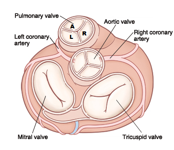
The pulmonary valve is a tricuspid valve is similar in construction and size of the aortic valve. The valve has a right (R) -, a left (L) - and an anterior slip valve (A). The pulmonary valve is slightly above, left anterior aortic valve. | The pulmonary valve is a tricuspid valve is similar in construction and size of the aortic valve. The valve has a right (R) -, a left (L) - and an anterior slip valve (A). The pulmonary valve is slightly above, left anterior aortic valve.<cite>2</cite> | ||
[[Image:Pulmvalv.png|400px]] | [[Image:Pulmvalv.png|400px]] | ||
Revision as of 23:11, 21 December 2013
Anatomy
The pulmonary valve is a tricuspid valve is similar in construction and size of the aortic valve. The valve has a right (R) -, a left (L) - and an anterior slip valve (A). The pulmonary valve is slightly above, left anterior aortic valve.[1]
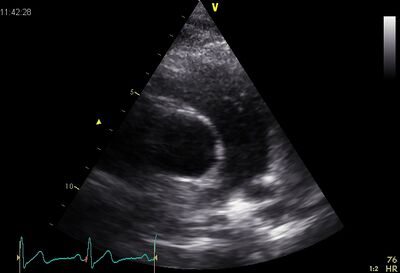
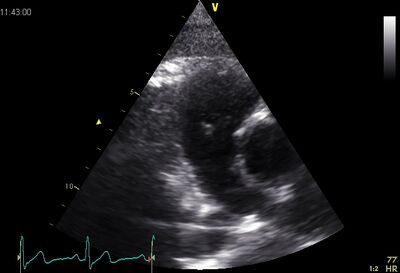
Echocardiographic views

|

|
| PSAX ao | Plax by tilted |
|---|
Stenosis
Stenosis of the pulmonary valve is very rare . Although as with aortic stenosis , the cause may be located above or below the valve , the valve usually is a wrong engineered valve . Which is then deformed dome formation with a small opening .
| Causes [1] | |
| Congenital | Tetralogy of Falot |
|---|---|
| Acquired |
|
Tetralogy of Fallot
Tetralogy of Fallot is a heart defect described by Etienne Fallot (1850-1911) , with four different heart defects occur:
- Pulmonaalstenose
- VSD
- RV Hypertrophy
- About propelled aortic
Tetralogy of Fallot results in a lower concentration of oxygen in the blood by the mixing of oxygen - rich blood and into the ventricles . The obstruction of the pulmonary valve ensures that blood from the right ventricle to the aorta flows through the ventricular septal defect . Usually this syndrome marked by cyanosis of the baby . This disorder was formerly called blue baby syndrome therefore called , but there are also " pink Fallots ", where the obstruction of the pulmonary valve is less severe . These patients are usually detected with an image of heart failure by excessive flow of the pulmonary vascular tree . In rare cases, there is a balanced position , wherein the stenosis is large enough in order to protect ( against over- flow) , and is low enough not to cause too much. Cyanosis pulmonary vascular
Click here for more info on Tetralogy of Fallot
Insufficiency
Pulmonaalinsufficiëntie is a volume load on the RV . Important insufficiency will lead to RV dilation. This volume will load the RV long endure , but will eventually go the RV failure.
| Causes [2] | |
| Physiologic | Is found in 40-80% of people |
|---|---|
| Congenital | Wrong landscaped valve cusps or absence of (partial) cover slip. |
| Acquired |
|
References
Error fetching PMID 20375260:
- Error fetching PMID 19065003:
- Error fetching PMID 20375260:
