Apical 2 chamber: Difference between revisions
| Line 34: | Line 34: | ||
==External links== | ==External links== | ||
* [https://www.techmed.sk/echo/ | * [https://www.techmed.sk/en/echo/view/20/ A2C - Image & video (TECHmED)] | ||
Latest revision as of 13:56, 9 January 2021
Content is incomplete and may be incorrect. |
| Author | I.A.C. van der Bilt | |
| Moderator | I.A.C. van der Bilt | |
| Supervisor | ||
| some notes about authorship | ||
How to Get a Good Apical Two Chamber View
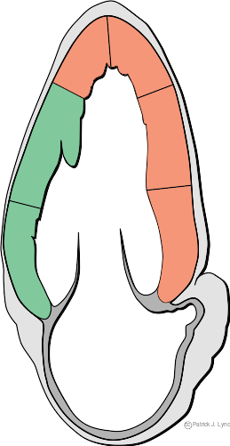
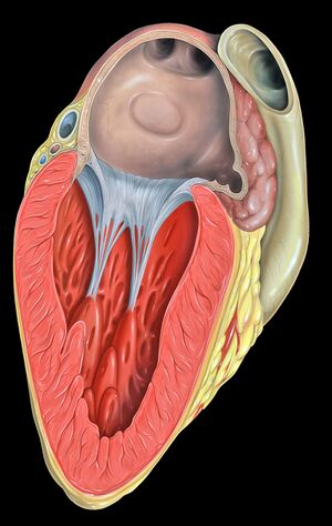
 The apical two chamber view is found by placing the transducer on the apex of the heart, near the Ictus Cordis. Rotation of the transducer by 35 to 45 degrees counterclockwise and angled toward the right shoulder will allow visualization of the apical two chamber view. This view permits the assessment of the inferior and anterior walls of the left ventricle. You will know that you have acquired a true apical two chamber view when right-sided structures are not seen. The landmark of a true apical two chamber is the coronary sinus, a small circular structure found at the atrioventricular junction.
Purposes of Getting a Good Apical Two Chamber View
This view permits the assessment and evaluation of left ventricular size and function. The coronary sinus, its size and structural condition may also be viewed. Its significance lies in the fact that a dilated coronary sinus can mean abnormal attachment of pulmonary veins and other abnormalities. Other pathologic processes such as the presence of inferior or anterior wall motion abnormalities can be seen using the apical two chamber view. Left ventricular thrombus can also be detected and its presence confirmed.
Doppler Imaging in Apical Two Chamber View
Doppler imaging is essentially done in this view to assess flow across the mitral valve. Patients without cardiac pathologies would manifest a flow pattern of two peaks. These are the E (rapid filling in the diastole) and the A (result of atrial contraction) waves. Normally, the A wave is smaller than the E wave. The presence of pathologies and the influence of other factors such as blood pressure and age, however, may cause different patterns. Color Doppler is used to appreciate flow direction and to determine velocity and pressure measurements.
Example of an Apical Two Chamber view
This a normal heart
| <flash>file=A2Cnormal.swf |
| Apical 2 chamber view of a normal heart |
