Aorta: Difference between revisions
No edit summary |
>Cvdw No edit summary |
||
| (7 intermediate revisions by 3 users not shown) | |||
| Line 1: | Line 1: | ||
==Aorta== | |||
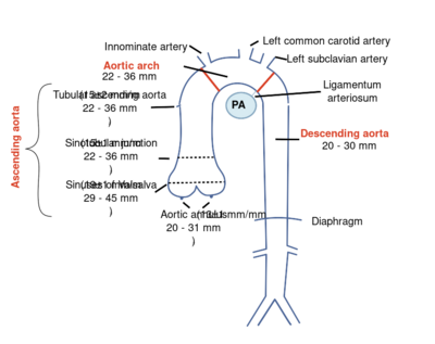
The thoracic aorta can be subdivided ito the aortic root (including the aortic annulus, aortic valve, and sinuses of Valsalva), the ascending aorta, the aortic arch, and the descending aorta. | |||
{| class="wikitable" cellpadding="0" cellspacing="0" border="0" width="600px" | |||
|- | |||
|bgcolor="#FFFFFF" align="center"|[[Image:Aortatract.svg|400px]] | |||
|- | |||
!Picture source: Eur J Echocardiogr 2010;11:645-58<cite>1</cite> | |||
|} | |||
==Aortic Dimensions== | ==Aortic Dimensions== | ||
Aortic dimensions decrease from sinuses of Valsalva to the descending aorta. | |||
{| class="wikitable" cellpadding="0" cellspacing="0" border="0" width=" | |||
{| class="wikitable" cellpadding="0" cellspacing="0" border="0" width="600px" | |||
|- | |- | ||
!colspan="2"|Aortic diameters (BSAindex) | !colspan="2"|Aortic diameters (BSAindex) | ||
|- | |- | ||
|Aortic annulus | !width="150px"|Aortic annulus | ||
|20 - 31mm (13 ± 1mm/m<sup>2</sup>) | |20 - 31mm (13 ± 1mm/m<sup>2</sup>) | ||
|- | |- | ||
!Aortic Root | |||
|29 - | |29 - 45 mm (19 ± 1 mm/m<sup>2</sup>) | ||
|- | |- | ||
!Sinotubular junction | |||
|22 - | |22 - 36 mm (15 ± 1 mm/m<sup>2</sup>) | ||
|- | |- | ||
!Tube | |||
|22 - | |22 - 36 mm (15 ± 2 mm/m<sup>2</sup>) | ||
|- | |- | ||
!Aortic Arch | |||
|22 - | |22 - 36 mm | ||
|- | |- | ||
!Descending aorta | |||
|20 - | |20 - 30 mm | ||
|- | |- | ||
!Abdominal aorta | |||
|20 - | |20 - 30 mm | ||
|- | |- | ||
|colspan="2"| | |colspan="2"| According to current recommendations measurements should be made using the leading edge to leading edge method, where callipers are placed on the outer layer of the anterior wall and the inner layer of the posterior wall. | ||
|} | |} | ||
==Aortic dissection== | ==Aortic dissection== | ||
Diagnostic is an undulating motion intimal flap , which in more recordings and directions must be seen. The flap should have a movement that is not parallel with any other cardio-thoracic structure . | Diagnostic is an undulating motion intimal flap, which in more recordings and directions must be seen. The flap should have a movement that is not parallel with any other cardio-thoracic structure. | ||
{| class="wikitable" cellpadding="0" cellspacing="0" border="0" width=" | {| class="wikitable" cellpadding="0" cellspacing="0" border="0" width="600px" | ||
|- | |- | ||
!valign="top"|Upon dissection watch: | !valign="top"|Upon dissection watch: | ||
| Line 56: | Line 60: | ||
|} | |} | ||
It also shows the intramural hematoma of the aorta to be aware of the aortic dissection. One variant This does not intraluminal flap was observed making the diagnosis is difficult to establish . Echocardiographic is viewed as a thickened aortic wall . | It also shows the intramural hematoma of the aorta to be aware of the aortic dissection. One variant This does not intraluminal flap was observed making the diagnosis is difficult to establish. Echocardiographic is viewed as a thickened aortic wall. | ||
Differentiation between true and false lumen: | {| class="wikitable" cellpadding="0" cellspacing="0" border="0" width="600px" | ||
|- | |||
!valign="top"|Differentiation between true and false lumen: | |||
| | |||
*In M mode, the flap moves to the false lumen in systole. | |||
*Spontaneous echo contrast and thrombus can be seen in the false lumen. | |||
*With color Doppler is delayed systolic flow seen by secondary or re-entry tear to the false lumen. | |||
*The false lumen (especially in chronic dissections) tends to be larger in comparison to the true lumen. | |||
|} | |||
==Aortic coarctation== | |||
Imaging of the aortic arch usually works best from the jugular (sternal supra). When evaluating a patient with a suspected coarctation always pay attention to associated anomalies such as: | Imaging of the aortic arch usually works best from the jugular (sternal supra). When evaluating a patient with a suspected coarctation always pay attention to associated anomalies such as: | ||
Determining coarctation | *Bicuspid aortic valve | ||
*Aortic valve stenosis | |||
Location Color doppler The | *Patent ductus arteriosus | ||
*VSD | |||
*Mitral valve abnormalities | |||
==Determining coarctation<cite>2</cite>== | |||
{| class="wikitable" cellpadding="0" cellspacing="0" border="0" width="600px" | |||
|- | |||
| | |||
!Instrument | |||
!Remark | |||
|- | |||
!width="100px"|Location | |||
|width="100px"|Color doppler | |||
|The origin of the carotid and subclavian artery are reference points for locating the coarctation. | |||
|- | |||
!Speed Profile | |||
|Continuous wave | |||
|Remember that collaterals systolic maximum speed but does reduce the diastolic gradient persists. In the presence of diastolic forward flow refers to a hemodynamically significant coarctation. | |||
Typical CW Doppler signal from descending aorta with diastolic forward flow matching hemodynamically significant coarctation. | |||
Typical CW Doppler signal from descending aorta with diastolic forward flow matching hemodynamically significant coarctation. | |} | ||
==References== | ==References== | ||
<biblio> | <biblio> | ||
#1 | #1 pmid=20823280 | ||
</biblio> | </biblio> | ||
Latest revision as of 10:43, 4 February 2016
Aorta
The thoracic aorta can be subdivided ito the aortic root (including the aortic annulus, aortic valve, and sinuses of Valsalva), the ascending aorta, the aortic arch, and the descending aorta.

|
| Picture source: Eur J Echocardiogr 2010;11:645-58[1] |
|---|
Aortic Dimensions
Aortic dimensions decrease from sinuses of Valsalva to the descending aorta.
| Aortic diameters (BSAindex) | |
|---|---|
| Aortic annulus | 20 - 31mm (13 ± 1mm/m2) |
| Aortic Root | 29 - 45 mm (19 ± 1 mm/m2) |
| Sinotubular junction | 22 - 36 mm (15 ± 1 mm/m2) |
| Tube | 22 - 36 mm (15 ± 2 mm/m2) |
| Aortic Arch | 22 - 36 mm |
| Descending aorta | 20 - 30 mm |
| Abdominal aorta | 20 - 30 mm |
| According to current recommendations measurements should be made using the leading edge to leading edge method, where callipers are placed on the outer layer of the anterior wall and the inner layer of the posterior wall. | |
Aortic dissection
Diagnostic is an undulating motion intimal flap, which in more recordings and directions must be seen. The flap should have a movement that is not parallel with any other cardio-thoracic structure.
| Upon dissection watch: |
|
|---|
It also shows the intramural hematoma of the aorta to be aware of the aortic dissection. One variant This does not intraluminal flap was observed making the diagnosis is difficult to establish. Echocardiographic is viewed as a thickened aortic wall.
| Differentiation between true and false lumen: |
|
|---|
Aortic coarctation
Imaging of the aortic arch usually works best from the jugular (sternal supra). When evaluating a patient with a suspected coarctation always pay attention to associated anomalies such as:
- Bicuspid aortic valve
- Aortic valve stenosis
- Patent ductus arteriosus
- VSD
- Mitral valve abnormalities
Determining coarctation[2]
| Instrument | Remark | |
|---|---|---|
| Location | Color doppler | The origin of the carotid and subclavian artery are reference points for locating the coarctation. |
| Speed Profile | Continuous wave | Remember that collaterals systolic maximum speed but does reduce the diastolic gradient persists. In the presence of diastolic forward flow refers to a hemodynamically significant coarctation.
Typical CW Doppler signal from descending aorta with diastolic forward flow matching hemodynamically significant coarctation. |
References
- Evangelista A, Flachskampf FA, Erbel R, Antonini-Canterin F, Vlachopoulos C, Rocchi G, Sicari R, Nihoyannopoulos P, Zamorano J, European Association of Echocardiography, Document Reviewers:, Pepi M, Breithardt OA, and Plonska-Gosciniak E. Echocardiography in aortic diseases: EAE recommendations for clinical practice. Eur J Echocardiogr. 2010 Sep;11(8):645-58. DOI:10.1093/ejechocard/jeq056 |