Normal Values of TEE: Difference between revisions

From Echopedia
Jump to navigation Jump to search
No edit summary
m (Reverted edits by 182.29.7.87 (talk) to last revision by April)
Tag: Rollback
 
(7 intermediate revisions by 3 users not shown)
Line 1: Line 1:
==Reference values for normal adult TEE measurements==
Reference values for normal adult TEE measurements. Adapted  from Perrino et al.<cite>Perrino</cite>
{| class="wikitable" style="font-size:90%;"


Line 108: Line 108:
<li><sup>d</sup>Left ventricular dimensions measured in transgastric mid short-axis view.</li>
<li>SD, standard deviation.</li>
<li>Adapted from Cohen G, White M, Sochowski R, et al. Reference values for normal transesophageal measurements. <em>J Am Soc Echocardiogr</em> 1995;8:221-230.</li>
<li>Adapted from Cohen et al. <cite>Cohen</cite></li>
</ul>
</ul>
|}
|}
Line 114: Line 114:
<!-- TABLE 3 -->
<!-- TABLE 3 -->


==Transesophageal Echocardiographic Anatomy==
{| class="wikitable" style="font-size:90%;"
|+'''Transesophageal Echocardiographic Anatomy'''
! ME two-chamber
| Probe adjustment: neutral
| Sector depth: ~14 cm
|-
| image
| valign="top" | Primary diagnostic issues
  <ul>
      <li>LA appendage</li>
      <li>Mass/thrombus</li>
      <li>LV apex pathology</li>
      <li>LV systolic dysfunction (apical segments)</li>
  </ul>
| valign="top" | Required structures
  <ul>
      <li>LA appendage</li>
      <li>Mitral valve</li>
      <li>LV apex (i.e., maximal LV length)</li>
  </ul>
|-
! ME LAX
| Probe adjustment: neutral
| Sector depth: ~12 cm
|-
| image
| valign="top" | Primary diagnostic issues
  <ul>
      <li>Mitral valve pathology</li>
      <li>LVOT pathology</li>
  </ul>
| valign="top" | Required structures
  <ul>
      <li>LV</li>
      <li>Mitral valve</li>
      <li>LVOT</li>
  </ul>
|-
! ME mitral commissural
| Probe adjustment: neutral
| Sector depth: ~12 cm
|-
| image
| valign="top" | Primary diagnostic issues
  <ul>
      <li>Localization of mitral valve pathology</li>
  </ul>
| valign="top" | Required structures
  <ul>
      <li>Mitral valve (P<sub>1</sub>, P<sub>3</sub> and A<sub>2</sub> scallops)</li>
      <li>Papillary muscles/chordae tendineae</li>
      <li>LA</li>
      <li>LV</li>
  </ul>
|-
! TG mid-SAX
| Probe adjustment: neutral
| Sector depth: ~12 cm
|-
| image
| valign="top" | Primary diagnostic issues
  <ul>
      <li>Hemodynamic instability</li>
      <li>LV enlargement</li>
      <li>LV hypertrophy</li>
      <li>LV systolic dysfunction (global and regional)</li>
  </ul>
| valign="top" | Required structures
  <ul>
      <li>LV cavity</li>
      <li>LV walls (at least 50% of circumference with visible endocardium)</li>
      <li>Papillary muscles (approximately equal in size and distinct from ventricular wall)</li>
  </ul>
|-
! TG two-chamber
| Probe adjustment: neutral
| Sector depth: ~12 cm
|-
| image
| valign="top" | Primary diagnostic issues
  <ul>
      <li>LV systolic dysfunction (anterior and inferior basal segments)</li>
  </ul>
| valign="top" | Required structures
  <ul>
      <li>Mitral leaflets</li>
      <li>Mitral subvalvular apparatus</li>
      <li>LV (anterior and inferior: basal plus mild segments)</li>
  </ul>
|-
! TG RV inflow
| Probe adjustment: neutral-rightward
| Sector depth: ~12 cm
|-
| image
| valign="top" | Primary diagnostic issues
  <ul>
      <li>RV systolic dysfunction</li>
      <li>Tricuspid valve pathology</li>
  </ul>
| valign="top" | Required structures
  <ul>
      <li>Tricuspid leaflets</li>
      <li>Tricuspid subvalvular apparatus</li>
  </ul>
|-
! TG RV inflow-outflow
| Probe adjustment: neutral-rightward
| Sector depth: ~14 cm
|-
| image
| valign="top" | Primary diagnostic issues
  <ul>
      <li>RV systolic dysfunction</li>
      <li>RVOT pathology</li>
      <li>Pulmonary artery pathology</li>
      <li>Pulmonic valve evaluation</li>
  </ul>
| valign="top" | Required structures
  <ul>
      <li>RA</li>
      <li>RV</li>
      <li>Main pulmonary artery</li>
      <li>Pulmonic valve</li>
  </ul>
|-
! TG basal SAX
| Probe adjustment: neutral
| Sector depth: ~12 cm
|-
| image
| valign="top" | Primary diagnostic issues
  <ul>
      <li>LV systolic dysfunction (basal segments)</li>
      <li>Mitral valve pathology</li>
  </ul>
| valign="top" | Required structures
  <ul>
      <li>Mitral leaflets</li>
      <li>Mitral subvalvular apparatus</li>
      <li>LV (basal segments)</li>
  </ul>
|-
! TG LAX
| Probe adjustment: neutral-leftward
| Sector depth: ~12 cm
|-
| image
| valign="top" | Primary diagnostic issues
  <ul>
      <li>LV systolic dysfunction (anteroseptal and posterior: basal segments)</li>
      <li>Doppler evaluation of aortic valve</li>
  </ul>
| valign="top" | Required structures
  <ul>
      <li>Mitral leaflets</li>
      <li>Mitral subvalvular apparatus</li>
      <li>LV (anteroseptal and posterior: basal plus midsegments)</li>
      <li>Aortic valve</li>
  </ul>
|-
! Deep TG LAX
| Probe adjustment: neutral
| Sector depth: ~16 cm
|-
| image
| valign="top" | Primary diagnostic issues
  <ul>
      <li>Aortic valve pathology</li>
      <li>LVOT pathology</li>
      <li>Doppler evaluation of aortic valve</li>
  </ul>
| valign="top" | Required structures
  <ul>
      <li>LV</li>
      <li>Aortic valve</li>
      <li>Aorta</li>
  </ul>
|-
| colspan="3" |
<ul>
<li>ME, midesophageal; Asc, ascending; SAX, short axis; LAX, long axis; UE, upper esophageal; Desc, descending; AV, aortic valve; RV, right ventricular; LVOT, left ventricular outflow tract; RA, right atrium; LA, left atrium; LV, left ventricular; RVOT, right ventricular outflow tract; TG, transgastric.</li>
<li>Modified from Miller JP, Lambert SA, Shapiro WA, et al. The adequacy of basic intraoperative transesophageal echocardiography performed by experienced anesthesiologist. <em>Anesth Analg</em> 2001;92:1103&ndash;1110, with permission.</li>
</ul>
|}
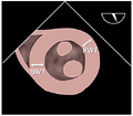
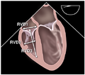
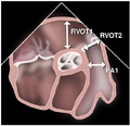
==Positions of measurements==
<gallery>
<gallery>
Line 331: Line 123:
File:fig009.svg
File:fig009.svg
</gallery>
</gallery>
==References==
<biblio>
#Perrino isbn=0781773296
#Cohen pmid=7640014
</biblio>

Latest revision as of 17:24, 17 October 2023

Reference values for normal adult TEE measurements. Adapted from Perrino et al.[1]

Reference values for normal adult TEE measurements
Parameter   Mean ± SD (mm) Range (mm)
  Right pulmonary artery diametera 17 ± 3 12-22
Left upper pulmonary vein diameter 11 ± 2 7-16
Left atrial appendage Length 28 ± 5 15-43
Diameter 16 ± 5 10-28
Superior vena cava diameter 15 ± 3 8-20
Right ventricular outflow tract diameterb 27 ± 4 16-36
Left atriumc Anteroposterior diameter 38 ± 6 20-52
Medial-lateral diameter 39 ± 7 24-52
Right atriumc Anteroposterior diameter 38 ± 5 28-52
Medial-lateral diameter 38 ± 6 29-53
Tricuspid annular diameterc 28 ± 5 20-40
Mitral annular diameterc 29 ± 4 20-38
Coronary sinus diameter 6.6 ± 1.5 4-10
Left ventricled Anteroposterior diameter (diastole) 43 ± 7 33-55
Medial-lateral diameter (diastole) 42 ± 7 23-54
Anteroposterior diameter (systole) 28 ± 6 18-40
Medial-lateral diameter (systole) 27 ± 6 18-42
Aortic root diameterb 28 ± 3 21-34
Descending thoracic aorta diameter Proximal 21 ± 4 14-30
Distal 20 ± 4 13-28
  • aRight pulmonary artery diameter measured in midesophageal ascending aorta short-axis view.
  • bAortic root and right ventricular outflow tract diameters measured in the midesophageal right ventricular inflow/outflow tract view.
  • cAtrial (end-systole) and both mitral and tricuspid annular (mid-diastole) diameters measured in the midesophageal four-chamber view.
  • dLeft ventricular dimensions measured in transgastric mid short-axis view.
  • SD, standard deviation.
  • Adapted from Cohen et al. [2]


Positions of measurements

References

  1. ISBN:0781773296 [Perrino]
  2. Cohen GI, White M, Sochowski RA, Klein AL, Bridge PD, Stewart WJ, and Chan KL. Reference values for normal adult transesophageal echocardiographic measurements. J Am Soc Echocardiogr. 1995 May-Jun;8(3):221-30. DOI:10.1016/s0894-7317(05)80031-8 | PubMed ID:7640014 | HubMed [Cohen]