TEE - standard imaging views: Difference between revisions
Jump to navigation
Jump to search
(Created page with ' ==Transesophageal Echocardiographic Anatomy== {| class="wikitable" style="font-size:90%;" |+'''Transesophageal Echocardiographic Anatomy''' ! ME two-chamber | Probe adjustment:...') |
m (moved Standard imaging views to TEE - standard imaging views) |
||
| (11 intermediate revisions by 2 users not shown) | |||
| Line 1: | Line 1: | ||
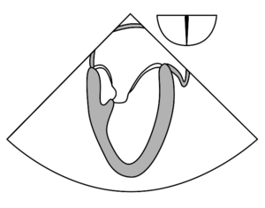
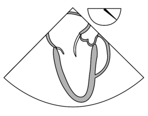
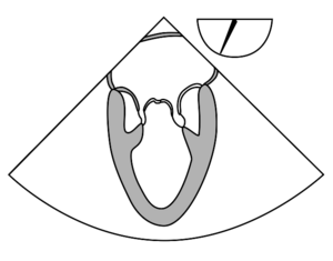
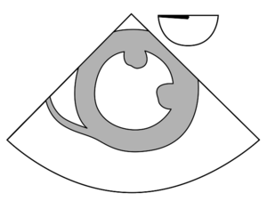
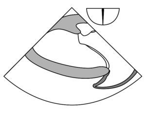
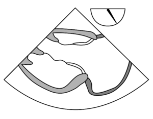
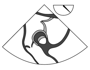
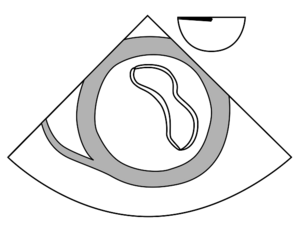
Transesophageal Echocardiographic imaging views. Adapted from Shanewise et al. <cite>Shanewise</cite> | |||
{| class="wikitable" style="font-size:90%;" | {| class="wikitable" style="font-size:90%;" | ||
| Line 9: | Line 9: | ||
|- | |- | ||
| | | [[File:ME_two-chamber.svg|center|thumb]] | ||
| valign="top" | Primary diagnostic issues | | valign="top" | Primary diagnostic issues | ||
<ul> | <ul> | ||
| Line 30: | Line 30: | ||
| Sector depth: ~12 cm | | Sector depth: ~12 cm | ||
|- | |- | ||
| | | [[File:ME_LAX.svg|center|thumb]] | ||
| valign="top" | Primary diagnostic issues | | valign="top" | Primary diagnostic issues | ||
<ul> | <ul> | ||
| Line 49: | Line 49: | ||
| Sector depth: ~12 cm | | Sector depth: ~12 cm | ||
|- | |- | ||
| | | [[File:ME_mitral_commissural.svg|center|thumb]] | ||
| valign="top" | Primary diagnostic issues | | valign="top" | Primary diagnostic issues | ||
<ul> | <ul> | ||
| Line 68: | Line 68: | ||
| Sector depth: ~12 cm | | Sector depth: ~12 cm | ||
|- | |- | ||
| | | [[File:TG_mid_SAX.svg|center|thumb]] | ||
| valign="top" | Primary diagnostic issues | | valign="top" | Primary diagnostic issues | ||
<ul> | <ul> | ||
| Line 89: | Line 89: | ||
| Sector depth: ~12 cm | | Sector depth: ~12 cm | ||
|- | |- | ||
| | | [[File:TG_two-chamber.svg|center|thumb]] | ||
| valign="top" | Primary diagnostic issues | | valign="top" | Primary diagnostic issues | ||
<ul> | <ul> | ||
| Line 107: | Line 107: | ||
| Sector depth: ~12 cm | | Sector depth: ~12 cm | ||
|- | |- | ||
| | | [[File:TG_RV_inflow.svg|center|thumb]] | ||
| valign="top" | Primary diagnostic issues | | valign="top" | Primary diagnostic issues | ||
<ul> | <ul> | ||
| Line 125: | Line 125: | ||
| Sector depth: ~14 cm | | Sector depth: ~14 cm | ||
|- | |- | ||
| | | [[File:TG_RV_inflow-outflow.svg|center|thumb]] | ||
| valign="top" | Primary diagnostic issues | | valign="top" | Primary diagnostic issues | ||
<ul> | <ul> | ||
| Line 147: | Line 147: | ||
| Sector depth: ~12 cm | | Sector depth: ~12 cm | ||
|- | |- | ||
| | | [[File:TG_basal_SAX.svg|center|thumb]] | ||
| valign="top" | Primary diagnostic issues | | valign="top" | Primary diagnostic issues | ||
<ul> | <ul> | ||
| Line 166: | Line 166: | ||
| Sector depth: ~12 cm | | Sector depth: ~12 cm | ||
|- | |- | ||
| | | [[File:TG_LAX.svg|center|thumb]] | ||
| valign="top" | Primary diagnostic issues | | valign="top" | Primary diagnostic issues | ||
<ul> | <ul> | ||
| Line 186: | Line 186: | ||
| Sector depth: ~16 cm | | Sector depth: ~16 cm | ||
|- | |- | ||
| | | [[File:Deep_TG_LAX.svg|center|thumb]] | ||
| valign="top" | Primary diagnostic issues | | valign="top" | Primary diagnostic issues | ||
<ul> | <ul> | ||
| Line 205: | Line 205: | ||
<ul> | <ul> | ||
<li>ME, midesophageal; Asc, ascending; SAX, short axis; LAX, long axis; UE, upper esophageal; Desc, descending; AV, aortic valve; RV, right ventricular; LVOT, left ventricular outflow tract; RA, right atrium; LA, left atrium; LV, left ventricular; RVOT, right ventricular outflow tract; TG, transgastric.</li> | <li>ME, midesophageal; Asc, ascending; SAX, short axis; LAX, long axis; UE, upper esophageal; Desc, descending; AV, aortic valve; RV, right ventricular; LVOT, left ventricular outflow tract; RA, right atrium; LA, left atrium; LV, left ventricular; RVOT, right ventricular outflow tract; TG, transgastric.</li> | ||
</ul> | </ul> | ||
|} | |} | ||
==References== | |||
<biblio> | |||
#Shanewise pmid=10512257 | |||
</biblio> | |||
Latest revision as of 13:11, 31 July 2010
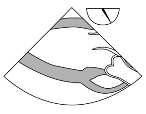
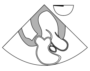
Transesophageal Echocardiographic imaging views. Adapted from Shanewise et al. [1]
| ME two-chamber | Probe adjustment: neutral | Sector depth: ~14 cm |
|---|---|---|
Primary diagnostic issues
|
Required structures
| |
| ME LAX | Probe adjustment: neutral | Sector depth: ~12 cm |
Primary diagnostic issues
|
Required structures
| |
| ME mitral commissural | Probe adjustment: neutral | Sector depth: ~12 cm |
Primary diagnostic issues
|
Required structures
| |
| TG mid-SAX | Probe adjustment: neutral | Sector depth: ~12 cm |
Primary diagnostic issues
|
Required structures
| |
| TG two-chamber | Probe adjustment: neutral | Sector depth: ~12 cm |
Primary diagnostic issues
|
Required structures
| |
| TG RV inflow | Probe adjustment: neutral-rightward | Sector depth: ~12 cm |
Primary diagnostic issues
|
Required structures
| |
| TG RV inflow-outflow | Probe adjustment: neutral-rightward | Sector depth: ~14 cm |
Primary diagnostic issues
|
Required structures
| |
| TG basal SAX | Probe adjustment: neutral | Sector depth: ~12 cm |
Primary diagnostic issues
|
Required structures
| |
| TG LAX | Probe adjustment: neutral-leftward | Sector depth: ~12 cm |
Primary diagnostic issues
|
Required structures
| |
| Deep TG LAX | Probe adjustment: neutral | Sector depth: ~16 cm |
Primary diagnostic issues
|
Required structures
| |
| ||
References
- Shanewise JS, Cheung AT, Aronson S, Stewart WJ, Weiss RL, Mark JB, Savage RM, Sears-Rogan P, Mathew JP, Quiñones MA, Cahalan MK, and Savino JS. ASE/SCA guidelines for performing a comprehensive intraoperative multiplane transesophageal echocardiography examination: recommendations of the American Society of Echocardiography Council for Intraoperative Echocardiography and the Society of Cardiovascular Anesthesiologists Task Force for Certification in Perioperative Transesophageal Echocardiography. Anesth Analg. 1999 Oct;89(4):870-84. DOI:10.1097/00000539-199910000-00010 |
