Case 4: Difference between revisions
Jump to navigation
Jump to search
Secretariat (talk | contribs) No edit summary |
Secretariat (talk | contribs) No edit summary |
||
| Line 18: | Line 18: | ||
|file_name5=E00109 | |file_name5=E00109 | ||
|descriptionfile5=Subcostal long axis showing pericardial effusion | |descriptionfile5=Subcostal long axis showing pericardial effusion | ||
|filepointer6=[[File:E00107.jpg| | |filepointer6=[[File:E00107.jpg|350px|left]] | ||
|file_name6= | |file_name6= | ||
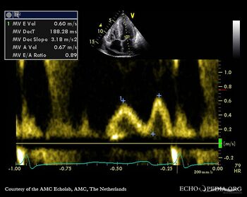
|descriptionfile6=Mitral valve inflow E<A a sign of [[diastolic dysfunction]] | |descriptionfile6=Mitral valve inflow E<A a sign of [[diastolic dysfunction]] | ||
}} | }} | ||
Latest revision as of 17:08, 15 July 2010
| Case description: This patient had amyloidosis with severe cardiac involvement | |
| Courtesy of: J. Vleugels, AMC, The Netherlands | |
| <flash>file=E00101.swf | <flash>file=E00102.swf |
| PLAX showing concentric left ventricular hypertrophy | PSAX shows severe concentric left and right ventricular hypertrophy, thickened mitral leaflets & pericardial effusion |
| <flash>file=E00103.swf | <flash>file=E00104.swf |
| PSAX through aortic valve which has thickened leaflets as well | A4CH showing severe left and right ventricular hypertrophy |
| <flash>file=E00109.swf | |
| Subcostal long axis showing pericardial effusion | Mitral valve inflow E<A a sign of diastolic dysfunction |
