Cardiac anatomy: Difference between revisions
No edit summary |
>Sonoquick (added a flow of structures to comment on and started with the right atrium) |
||
| Line 1: | Line 1: | ||
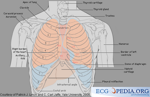
For understanding and interpreting echocardiograms, sound anatomical knowledge is invaluable. [[Image:Torso.png|thumb| Structures in the chest]] | For understanding and interpreting echocardiograms, sound anatomical knowledge is invaluable. | ||
The primary function of the heart must be adressed prior to breaking down the individual cardiac structures and their respective physiological properties. Each organ system in the body is important, but without the heart, none of them would function, whether alone or in unison. The heart is the body's life pump or motor, responsible for pumping blood throughout the cardiovascular system. The blood pumped by the heart helps heat and cool the body and allows oxygen/carbon dioxide gas exchange at both the cellular level and in the lung tissue. The pressure that the heart creates, in tandem with the arterial vessels, shuttles blood through the major organs, cleaning the blood and allowing the body to fight infection. | |||
And now for THE most important organ in the body... | |||
Let's track the blood through the heart from the point of entry. | |||
Right Atrium | |||
The right atrium, the right upper chamber of the heart, has many important functions. First, it is the receptacle for all de-oxygenated blood returning to the heart via various vessels. The Inferior Vena Cava (IVC) and Superior Vena Cava (SVC) return blood from the lower body and upper body/head into the medial aspect of the heart. The coronary sinus (CS) vein and thebesian vessels carry the used blood from the coronary artery system that supplies the heart muscle with it's own blood. The right atrium also has a small pouch-like tissue sac called an atrial appendage. The right atrium also houses the beginning of the heart's self-powered electrical system. The Sino-atrial node ("SA Node") sends and electrical impulse down a pathway that will be discussed in length later in cardiac anatomy. The right atrium is adjacent to the left atrium and is usually completely isolated from it by a thin layer of tissue called the atrial septum. Just like the septum in your nose, it keeps the right and left sides separate, but in the presence of a defect blood can cross over from one side to the other. Right in the middle of septum, the the tissue gets very, very thin. This thin spot is called the fossa ovalis. In utero, a fetus does not breathe air to absorb oxygen, so blood does not get pumped to the lungs. the blood is shuttled through a hole, purposefully placed by nature, in the atrial septum from the right atrium to the left atrium. This allows the blood to bypass the lungs and go to the left heart and be pumped back to the mother via the umbilical cord. Upon birth, this hole usuall closes completely or develops a flap that keeps tight by the immense pressure the left heart places upon it. the right atrium may also allow you to visualize a thin network of string-like structures called the chiari network. This appears during echocardiography as thin wavy filaments that undulate with the bloodflow. | |||
Tricuspid Valve | |||
Right Ventricle | |||
Pulmonic Valve | |||
Pulmonary Artery | |||
Pulomnary Veins | |||
Left Atrium | |||
Mitral Valve (Bicuspid Valve) | |||
Left Ventricle | |||
Aortic Valve | |||
Aorta | |||
Arterial Vasculature | |||
Venous System | |||
Cardiac Electrical System | |||
[[Image:Torso.png|thumb| Structures in the chest]] | |||
Revision as of 04:41, 5 February 2009
For understanding and interpreting echocardiograms, sound anatomical knowledge is invaluable.
The primary function of the heart must be adressed prior to breaking down the individual cardiac structures and their respective physiological properties. Each organ system in the body is important, but without the heart, none of them would function, whether alone or in unison. The heart is the body's life pump or motor, responsible for pumping blood throughout the cardiovascular system. The blood pumped by the heart helps heat and cool the body and allows oxygen/carbon dioxide gas exchange at both the cellular level and in the lung tissue. The pressure that the heart creates, in tandem with the arterial vessels, shuttles blood through the major organs, cleaning the blood and allowing the body to fight infection.
And now for THE most important organ in the body...
Let's track the blood through the heart from the point of entry.
Right Atrium
The right atrium, the right upper chamber of the heart, has many important functions. First, it is the receptacle for all de-oxygenated blood returning to the heart via various vessels. The Inferior Vena Cava (IVC) and Superior Vena Cava (SVC) return blood from the lower body and upper body/head into the medial aspect of the heart. The coronary sinus (CS) vein and thebesian vessels carry the used blood from the coronary artery system that supplies the heart muscle with it's own blood. The right atrium also has a small pouch-like tissue sac called an atrial appendage. The right atrium also houses the beginning of the heart's self-powered electrical system. The Sino-atrial node ("SA Node") sends and electrical impulse down a pathway that will be discussed in length later in cardiac anatomy. The right atrium is adjacent to the left atrium and is usually completely isolated from it by a thin layer of tissue called the atrial septum. Just like the septum in your nose, it keeps the right and left sides separate, but in the presence of a defect blood can cross over from one side to the other. Right in the middle of septum, the the tissue gets very, very thin. This thin spot is called the fossa ovalis. In utero, a fetus does not breathe air to absorb oxygen, so blood does not get pumped to the lungs. the blood is shuttled through a hole, purposefully placed by nature, in the atrial septum from the right atrium to the left atrium. This allows the blood to bypass the lungs and go to the left heart and be pumped back to the mother via the umbilical cord. Upon birth, this hole usuall closes completely or develops a flap that keeps tight by the immense pressure the left heart places upon it. the right atrium may also allow you to visualize a thin network of string-like structures called the chiari network. This appears during echocardiography as thin wavy filaments that undulate with the bloodflow.
Tricuspid Valve
Right Ventricle
Pulmonic Valve
Pulmonary Artery
Pulomnary Veins
Left Atrium
Mitral Valve (Bicuspid Valve)
Left Ventricle
Aortic Valve
Aorta
Arterial Vasculature
Venous System
Cardiac Electrical System
