Apical 2 chamber: Difference between revisions
Jump to navigation
Jump to search
No edit summary |
No edit summary |
||
| Line 9: | Line 9: | ||
|file_name=A2Cnormal | |file_name=A2Cnormal | ||
}} | }} | ||
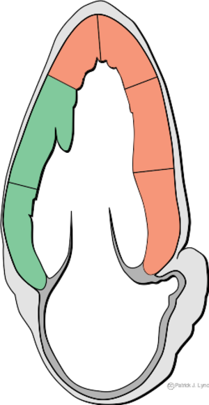
Image:Heart_apical_2c_myocardial_regions.svg | [[Image:Heart_apical_2c_myocardial_regions.svg|thumb|Image showing the left parasternal long axis transection (PSLAX) of the heart by the ultrasound waves]] | ||
{{clr}} | {{clr}} | ||
Revision as of 22:30, 16 June 2008
Content is incomplete and may be incorrect. |
The apical two chamber view is found by placing the transducer on the apex of the heart, near the Ictus Cordis.
Example of an apical 2 chamber view
This a normal heart
| <flash>file=A2Cnormal.swf |
| Apical 2 chamber view of a normal heart |
