Dobutamine Stress Echo
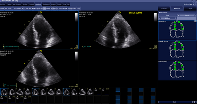
DSE to assess regional wall motion disorders A stress echo is a study whereby the function of the LV is at rest and during exercise. The heart is charged by means of dobutamine intravenously. Dobutamine is a catecholamine with predominant β - receptor stimulator which positive inotropic, chronotropic and dromotropic effects caused by the heart. Dobutamine is the half-life of approximately 2 minutes. The inotropic and chronotropic effects induce myocardial ischemia in a significant narrowing of a coronary artery. Echocardiography than regional wall motion detect disorders. DSE is also used to detect contractile reserve on myocardial damage after infarction. The betrouwbaardheid for detecting coronary stenosis can be compared with myocardial perfusion scintigraphy and DSE is superior to exercise ECG.
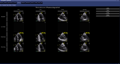
Advance is first made with the echo resting platelets : A Plax, PSax pm, AP4Ch and AP2Ch. During the run of Dobutamine the heart is properly monitored using a 12 -channel ECG and blood pressure. With each increase in dose dobutamine (every 3 minutes see table) is an ECG and blood pressure measured. The research is safe, but can cause symptoms such as: palpitations, chest pain, dizzy or light present in the head. The Dobutamine is stopped when reaching the maximum heart rate [(220 - age) x 0.85], severe complications or incidents, demonstrable new wandbewegingstoonissen in more than one segment or an increase in end- systolic volume. In some cases such as heart rate hetmaximale not yet been reached, there may be also 0.25mg atropine be co-administered with a maximum of 1 mg (at intervals of 1 minute). Next Dobutamine
At maximum heart rate are again echo recordings. Another Plax, PSax pm, AP4Ch and AP2Ch, exactly like the rest plates (otherwise do not compare the recordings with each other) that requires only routine of the sonographer. Then ensure that the heart rate is slow again.[1]

|

|
| DSE assessment on RWBS | DSE protocol |
|---|
| Relationship dobutamine concentration by weight and pump mode. Dobutamine 250mg in 50ml, pump speed in ml / h. (Copyright © OLVG) | |||||
|---|---|---|---|---|---|
| Time in minutes | |||||
| 0 | 0 | 3 | 6 | 9 | |
| Dobutamine in µg/kg/min | |||||
| 5 | 10 | 20 | 30 | 40 | |
| Weight in kg | Pump Stand | ||||
| 44 | 2.6 | 5.3 | 10.5 | 15.7 | 20.9 |
| 46 | 2.8 | 5.6 | 11.2 | 16.8 | 22.4 |
| 48 | 2.9 | 5.8 | 11.6 | 17.4 | 23.2 |
| 50 | 3.0 | 6.0 | 12.0 | 18.0 | 24.0 |
| 52 | 3.1 | 6.2 | 12.4 | 18.6 | 24.8 |
| 54 | 3.2 | 6.5 | 13.0 | 19.4 | 25.6 |
| 56 | 3.4 | 6.8 | 13.6 | 20.4 | 27.2 |
| 58 | 3.5 | 7.0 | 14.0 | 21.0 | 28.0 |
| 60 | 3.6 | 7.2 | 14.4 | 21.6 | 28.8 |
| 62 | 3.7 | 7.4 | 14.8 | 22.2 | 28.8 |
| 64 | 3.8 | 7.6 | 15.2 | 22.8 | 30.4 |
| 66 | 4.0 | 8.0 | 16.0 | 24.0 | 32.0 |
| 68 | 4.1 | 8.2 | 16.4 | 24.6 | 32.8 |
| 70 | 4.2 | 8.4 | 16.8 | 25.2 | 33.6 |
| 72 | 4.3 | 8.6 | 17.2 | 25.8 | 34.4 |
| 74 | 4.4 | 8.8 | 17.6 | 26.4 | 35.2 |
| 76 | 4.6 | 9.2 | 18.4 | 27.6 | 36.8 |
| 78 | 4.7 | 9.4 | 18.8 | 28.2 | 37.6 |
| 80 | 4.8 | 9.6 | 19.2 | 28.8 | 38.4 |
| 82 | 4.9 | 9.8 | 19.6 | 29.4 | 39.2 |
| 84 | 5.0 | 10.0 | 20.0 | 30.0 | 40.0 |
| 86 | 5.1 | 10.2 | 20.4 | 30.6 | 40.8 |
| 88 | 5.3 | 10.6 | 21.2 | 31.8 | 42.4 |
| 90 | 5.4 | 10.8 | 21.6 | 32.4 | 43.2 |
| 92 | 5.5 | 11.0 | 22.0 | 33.0 | 44.0 |
| 94 | 5.6 | 11.2 | 22.4 | 33.6 | 44.8 |
| 96 | 5.8 | 11.6 | 23.2 | 34.8 | 46.4 |
| 98 | 5.9 | 11.8 | 23.6 | 35.4 | 47.2 |
| 100 | 6.0 | 12.0 | 24.0 | 36.0 | 48.0 |
| 102 | 6.1 | 12.2 | 24.4 | 36.6 | 48.4 |
| 104 | 6.2 | 12.4 | 24.8 | 37.2 | 49.6 |
| 106 | 6.4 | 12.8 | 25.6 | 38.4 | 51.2 |
| 108 | 6.5 | 13.0 | 26.0 | 39.0 | 52.0 |
| 110 | 6.6 | 13.2 | 26.4 | 39.6 | 52.8 |
DSE with low- gradient aortic stenosis Low- gradient aortic stenosis is defined as a severe aortic valve stenosis (AVA < 1.0cm ²) with a transvalvular pressure gradient ≤ 30mmHg. A low- gradient aortic stenosis occurs in patients who have LV dysfunction with decreased ejection fraction. The assessment of the AGM in these patients may be overestimated because the calculated AVA is proportional to the displacement. A poorly functioning LV can exert enough pressure to open. Calcified aortic valve sufficient At a fixed, or "true " stenosis in which the stroke volume increases, the gradient across the valve is relatively also increases. In some patients, an increase in stroke volume results for only a limited increase in pressure gradient across the aortic valve. This phenomenon is called a " pseudo stenosis ". DSE is a tool to distinguish between a true and a pseudo stenosis stenosis. Distinction
Patients with pseudo- stenosis manifest an increase in the calculated AGM and a decrease in resistance of the valve as a response to an increase of the stroke volume. The reaction is different in patients with severe aortic stenosis true in whom a dobutamine - induced increase in the transvalvular flow, giving an increase in the mean transvalvular gradient, but no change in AGM.
A DSE study in a low- gradient aortic valve stenosis is given a low -dose dobutamine. That is, they started to get 5μg/kg/min and gradually increased to a maximum 20μg/kg/min.
If there is an increase in stroke volume, an increase in AVA ≥ 0.3cm ² and a small change in gradient produced after administration of Dobutamine there is an overestimation of the severity of aortic stenosis (= pseudo stenosis).
A DSE may also demonstrate a severe aortic stenosis with low transvalvular pressure gradient or contractile reserve. Contractile reserve has a predictive value for mortality in surgery of aortic valve replacement. Recent studies have shown that perioperative mortality 5-8 % versus 32 % without contractile reserve in contractile reserve.[2]
Indication DSE
| Indications for DSE |
|---|
| Suspected coronary artery disease |
| Inability to perform bicycle test |
| Known for determining coronary ischemia before and after revascularization |
| Known coronary artery disease to determine areas of ischemia |
| Preoperatively, to assess risk, with large myocardial infarction. |
| Detection of viability |
Contra-indication DSE
| Contra-indications for DSE |
|---|
| Acute myocardial infarction (≤ 4-10 days) |
| Unstable angina |
| Known relevant main stem stenosis |
| Congestive heart failure |
| Serious tachyarrhytmiën |
| Serious klepstenosen |
| Hypertrophic obstructive cardiomyopathy |
| Acute pericarditis, myocarditis, endocarditis |
| Aortic dissection |
References
- Krahwinkel W, Ketteler T, Gödke J, Wolfertz J, Ulbricht LJ, Krakau I, and Gülker H. Dobutamine stress echocardiography. Eur Heart J. 1997 Jun;18 Suppl D:D9-15. DOI:10.1093/eurheartj/18.suppl_d.9 |
- Lange RA and Hillis LD. Dobutamine stress echocardiography in patients with low-gradient aortic stenosis. Circulation. 2006 Apr 11;113(14):1718-20. DOI:10.1161/CIRCULATIONAHA.105.617159 |