Diastolic Function
Jump to navigation
Jump to search
The Left Ventricle
There is still much uncertainty about the pathophysiology of diastolic heart failure, which still lacks an effective treatment, with a high mortality related to diastolic heart failure over the last decades. Diastolic heart failure is characterized by an increased LV diastolic stiffness. Echocardiographic there are some important measurements are indispensable to good to estimate. Stiffness in the standard ultrasound examination.
Left ventricular diastolic function
Normal Values diastolic parameters | |||||
|---|---|---|---|---|---|
| Measurement | Age group (y) | ||||
| 16-20 | 21-40 | 41-60 | >60 | ||
| IVRT (ms) | 50 ± 9 (32-68) | 67 ± 8 (51-83) | 74 ± 7 (60-88) | 87 ± 7 (73-101) | |
| E/A ratio | 1.88 ± 0.45 (0.98-2.78) | 1.53 ± 0.40 (0.73-2.33) | 1.28 ± 0.25 (0.78-1.78) | 0.96 ± 0.18 (0.6-1.32) | |
| DT (ms) | 142 ± 19 (104-180) | 166 ± 14 (138-194) | 181 ± 19 (143-219) | 200 ± 29 (142-258) | |
| A duration (ms) | 113 ± 17 (79-147) | 127 ± 13 (101-153) | 133 ± 13 (107-159) | 138 ± 19 (100-176) | |
| PV S/D ratio | 0.82 ± 0.18 (0.46-1.18) | 0.98 ± 0.32 (0.34-1.62) | 1.21 ± 0.2 (0.81-1.61) | 1.39 ± 0.47 (0.45-2.33) | |
| PV Ar (cm/s) | 16 ± 10 (1-36) | 21 ± 8 (5-37) | 23 ± 3 (17-29) | 25 ± 9 (11-39) | |
| PV Ar duration (ms) | 66 ± 39 (1-144) | 96 ± 33 (30-162) | 112 ± 15 (82-142) | 113 ± 30 (53-173) | |
| Septal e´ (cm/s) | 14.9 ± 2.4 (10.1-19.7) | 15.5 ± 2.7 (10.1-20.9) | 12.2 ± 2.3 (7.6-16.8) | 10.4 ± 2.1 (6.2-14.6) | |
| Septal e´/a´ ratio | 2.4* | 1.6 ± 0.5 (0.6-2.6) | 1.1 ± 0.3 (0.5-1.7) | 0.85 ± 0.2 (0.45-1.25) | |
| Lateral e´ (cm/s) | 20.6 ± 3.8 (13-28.2) | 19.8 ± 2.9 (14-25.6) | 16.1 ± 2.3 (11.5-20.7) | 12.9 ± 3.5 (5.9-19.9) | |
| Lateral e´/a´ ratio | 3.1* | 1.9 ± 0.6 (0.7-3.1) | 1.5 ± 0.5 (0.5-2.5) | 0.9 ± 0.4 (0.1-1.7) | |
| |||||
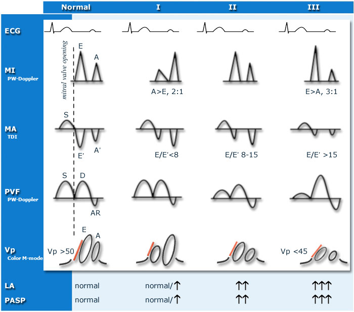
Schematic diastolic filling patterns |
|---|
| A patient with dyspnea, preserved systolic LV function, dilated left atrium and elevated pulmonary artery systolic pressure, without any significant mitral valve disease that could explain these findings, is the patient that requires an intensified search for diastolic LV dysfunction. |

|
| I: impaired relaxation, II: moderate diastolic dysfunction (pseudonormal), III: restrictive left ventricular filling (impaired LV compliance), ECG: electrocardiogram, MI: mitral inflow, MA: mitral annular velocities, PVF: pulmonary venous flow, Vp: velocity of flow progression, LA: left atrium, PASP: pulmonary artery systolic pressure.[2]
Click here for animation on diastolic dysfunction |
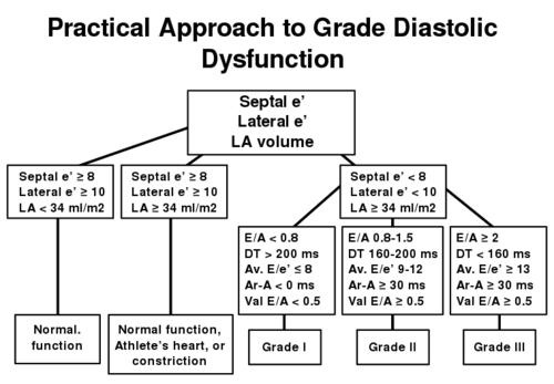
Diastolic function flowchart [1] |
|---|

|