Mitral Valve
Anatomy
The mitral valve consists of two valve leaflets, the anterior leaflet (AMVL) and the posterior leaflet (PMVL), which together have a surface of 4 - 6 cm2. Via chordae tendineae, small tendons which ensure that the leaflets do not prolapse, the valve leaflets are attached to two major papillary muscles (anterolateral en posteromedial) in the left ventricle.
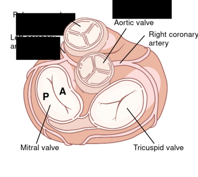
|
| Top view of the normal heart showing the coronary arteries and four valves including the mitral valve with its anterior (A) and posterior (P) leaflets1 |
|---|
The mitral valve is bringing many scan faces in pictures: PLAX, PSAXmv, AP4Ch, AP5Ch, AP2Ch, AP3Ch and subcostal4Ch. Mitral valvular insufficiency should be considered in all views. When significant abnormalities of the mitral valve are suspected, TEE (particularly 3D TEE) can assist in assessing the severity.
| Video |
| 3D (TEE) view of MV with a chorda rupture |
|---|
| Examples of Mitral variants (AP4Ch) | |
|---|---|
| Video | Video |
| Normal | Degenerative |
| Video | Video |
| Prolapse | Rheumatic |
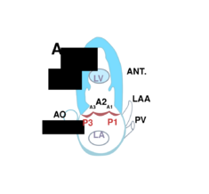
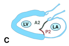
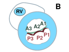
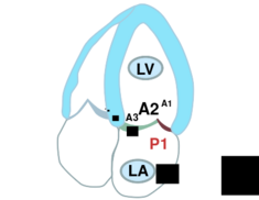
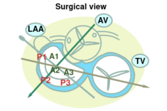
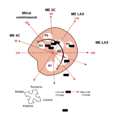
The scallops of the mitral valve
The two leaflets are divided into a total of six scallops: A1, A2, A3 (anterior) and P1, P2, P3 (posterior).
Stenosis
| Causes of mitral valve stenosis | |
|---|---|
| Acquired |
|
| Space occupying lesion |
|
| Congenital |
|
Effects of mitral valve stenosis:
Chest pain, of breath when lying flat, with exertion and attacks during the night or morning (cardiac asthma), possibly coughing up blood; any complaints from right valve failure, such as ankle edema. Palpitations by atrial fibrillation, which can cause embolism, such as a stroke.
Treatment
The valve can be opened by balloon angioplasty or surgically treated by valve replacement.
Click here for mitral stenosis quantification.
Insufficiency
| Causes of mitral valvular insufficiency | |
|---|---|
| Annular dilatation |
|
| Degenerative disease |
|
| Acquired valve defect |
|
| Secondary |
|
Mitral valve insufficiency (MI) may result in left-sided congestive heart failure. An MI results in back flow of blood from the left ventricle into the left atrium. This may eventually result in pulmonary hypertension and pulmonary edema. Pulmonary edema is manifested by shortness of breath, initially with exertion, but later at rest, orthopnea and nocturnal dyspnea attacks. On physical examination, pulmonary edema can be detected by fine crackles, particularly in the dorsobasale lung fields. Increasing left atrial pressure and dimension may cause atrial fibrillation.
Click here for quantification mitral valvular insufficiency .
References
<biblio>
- 1 How the heart works from Geisinger’s Health Library.
- 2 Mechanisms of Mitral regugitation - ML Geleijnse.
</biblio>
