| Subvalvular membrane
|
|
|
| Courtesy of: AMC Echolab, AMC, The Netherlands
|
|
|
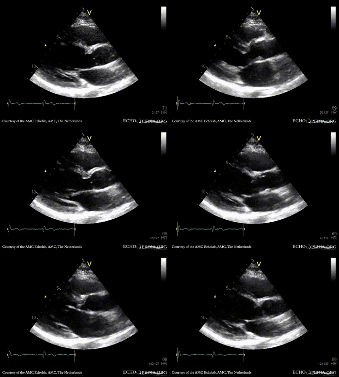
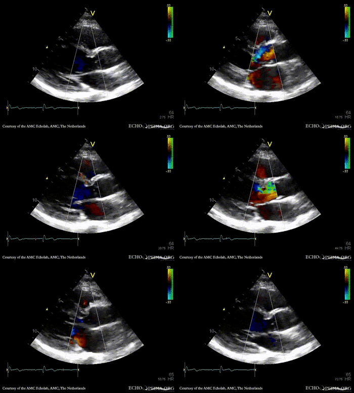
| PSAX: subvalvular membrane
|
PSAX with Color Doppler: high velocity flow in LVOT
|
 enlarge enlarge
 download file to use in your powerpoint presentation download file to use in your powerpoint presentation
|
 enlarge enlarge
 download file to use in your powerpoint presentation download file to use in your powerpoint presentation
|
|
|
|
|
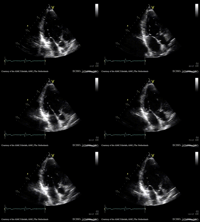
| A3CH: subvalvular membrane
|
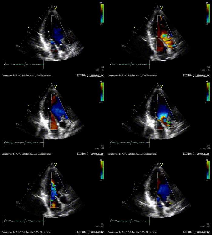
A3CH with Color Doppler: high velocity flow in LVOT
|
 enlarge enlarge
 download file to use in your powerpoint presentation download file to use in your powerpoint presentation
|
 enlarge enlarge
 download file to use in your powerpoint presentation download file to use in your powerpoint presentation
|
|
|
|
|
|
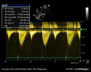
| Continuous-wave Doppler signal of transaortic flow
|
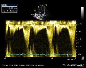
Pulsed-wave Doppler signal of LVOT flow: mild dynamic gradient
|
|
|
|
|
|
|
|
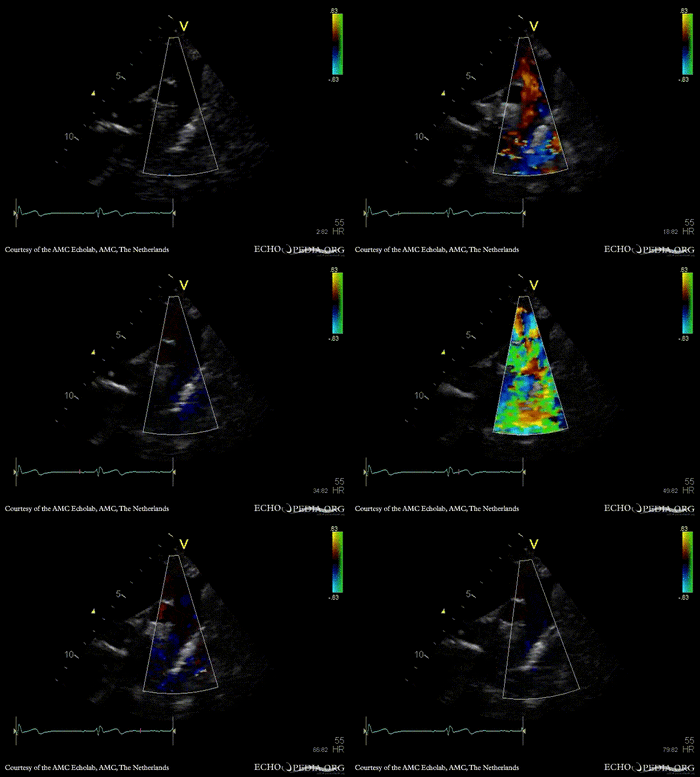
| Suprasternal view: high velocity flow in ascending aorta
|
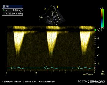
Continuous-wave Doppler signal of flow in ascending aorta
|
 enlarge enlarge
 download file to use in your powerpoint presentation download file to use in your powerpoint presentation
|
|
|
|
|
|
|
|
|
|
|
|
|


