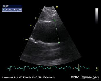
| Dilated aortic root and aortic regurgitation
|
|
|
| Courtesy of: AMC Echolab, AMC, The Netherlands
|
| <html5media height="350" width="279" autoplay="true" loop="true">File:E00236.mp4</html5media>
|
|
| PLAX: dilated aortic root
|
PLAX: dilated ascending aorta
|
 enlarge enlarge
 download file to use in your powerpoint presentation download file to use in your powerpoint presentation
|
|
|
|
| <html5media height="350" width="279" autoplay="true" loop="true">File:E00238.mp4</html5media>
|
<html5media height="350" width="279" autoplay="true" loop="true">File:E00239.mp4</html5media>
|
| A4CH: dilated left ventricle, false tendon in the apex and Chiari network in the right atrium
|
A3CH: Color Doppler, severe aortic regurgitation, eccentric jet
|
 enlarge enlarge
 download file to use in your powerpoint presentation download file to use in your powerpoint presentation
|
 enlarge enlarge
 download file to use in your powerpoint presentation download file to use in your powerpoint presentation
|
|
|
| <html5media height="350" width="279" autoplay="true" loop="true">File:E00240.mp4</html5media>
|
<html5media height="350" width="279" autoplay="true" loop="true">File:E00241.mp4</html5media>
|
| A5CH: Color Doppler, severe aortic regurgitation, eccentric jet
|
A5CH: dilated aortic root
|
 enlarge enlarge
 download file to use in your powerpoint presentation download file to use in your powerpoint presentation
|
 enlarge enlarge
 download file to use in your powerpoint presentation download file to use in your powerpoint presentation
|
|
|
| <html5media height="350" width="279" autoplay="true" loop="true">File:E00242.mp4</html5media>
|
|
| Subcostal view: severe aortic regurgitation, eccentric jet
|
|
 enlarge enlarge
 download file to use in your powerpoint presentation download file to use in your powerpoint presentation
|
|
|
|
|
|
|
|
|
|
|
|
|
