Case 177

From Echopedia
Revision as of 20:13, 1 December 2023 by Zoran (talk | contribs) (Replace html5media with gif)
(diff) ← Older revision | Latest revision (diff) | Newer revision → (diff)
Jump to navigation Jump to search
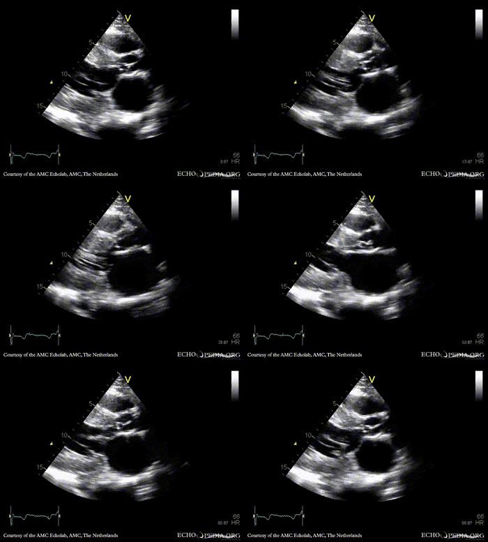
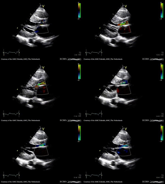
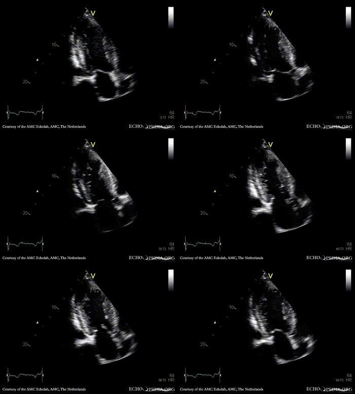
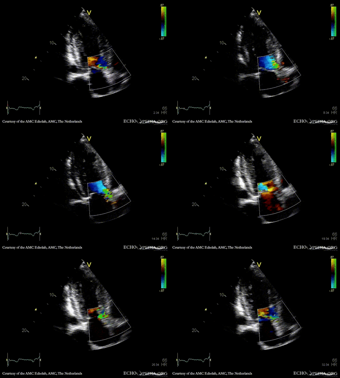
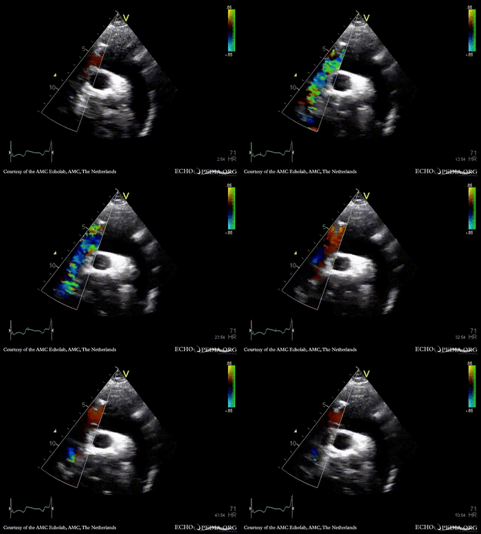
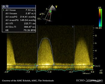
Subvalvular membrane in left ventricle outflow tract
Courtesy of: AMC Echolab, AMC, The Netherlands
E00911.gif E00912.gif
PLAX: subvalvular membrane in left ventricle outflow tract PLAX with Color Doppler: high velocity flow in left ventricle outflow tract
Magnifier.pngenlarge

Ppt logo.pngdownload file to use in your powerpoint presentation

Magnifier.pngenlarge

Ppt logo.pngdownload file to use in your powerpoint presentation

E00913.gif E00914.gif
A3CH A3CH with Color Doppler
Magnifier.pngenlarge

Ppt logo.pngdownload file to use in your powerpoint presentation

Magnifier.pngenlarge

Ppt logo.pngdownload file to use in your powerpoint presentation

E00915.gif
E00917.jpg
Suprasternal view: Collor Doppler, high velocity flow in left ventricle outflow tract Continuous-wave signal of high flow in left ventricle outflow tract
Magnifier.pngenlarge

Ppt logo.pngdownload file to use in your powerpoint presentation