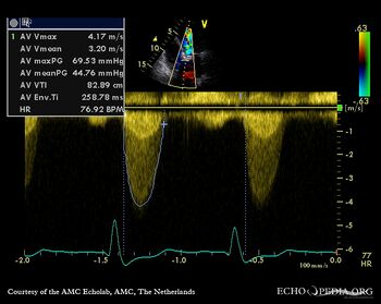
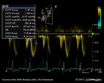
| Severe aortic valve stenosis and plaque in abdominal aorta
|
|
|
| Courtesy of: AMC Echolab, AMC, The Netherlands
|
| <html5media height="350" width="279" autoplay="true" loop="true">File:E00772.mp4</html5media>
|
<html5media height="350" width="279" autoplay="true" loop="true">File:E00773.mp4</html5media>
|
| PLAX: concentric hypertrophy of left ventricle, thickend aortic valve
|
PSAX: concentric hypertrophy of left ventricle
|
 enlarge enlarge
 download file to use in your powerpoint presentation download file to use in your powerpoint presentation
|
 enlarge enlarge
 download file to use in your powerpoint presentation download file to use in your powerpoint presentation
|
|
|
| <html5media height="350" width="279" autoplay="true" loop="true">File:E00774.mp4</html5media>
|
<html5media height="350" width="279" autoplay="true" loop="true">File:E00775.mp4</html5media>
|
| PSAX: stenosis of aortic valve
|
PLAX with Color Doppler: high velocity transaortic flow, mild aortic regurgitation
|
 enlarge enlarge
 download file to use in your powerpoint presentation download file to use in your powerpoint presentation
|
 enlarge enlarge
 download file to use in your powerpoint presentation download file to use in your powerpoint presentation
|
|
|
| <html5media height="350" width="279" autoplay="true" loop="true">File:E00776.mp4</html5media>
|
<html5media height="350" width="279" autoplay="true" loop="true">File:E00777.mp4</html5media>
|
| A4CH: concentric hypertrophy of left ventricle
|
A3CH with Color Doppler: high velocity transaortic flow, mild aortic regurgitation
|
 enlarge enlarge
 download file to use in your powerpoint presentation download file to use in your powerpoint presentation
|
 enlarge enlarge
 download file to use in your powerpoint presentation download file to use in your powerpoint presentation
|
|
|
|
|
|
| Continuous-wave Doppler signal of transaortic flow
|
Pulsed-wave signal of flow in LVOT
|
|
|
|
|
|
| <html5media height="350" width="279" autoplay="true" loop="true">File:E00780.mp4</html5media>
|
<html5media height="350" width="279" autoplay="true" loop="true">File:E00781.mp4</html5media>
|
| Subcostal view: plaque in abdominal aorta
|
Subcostal view: plaque in abdominal aorta
|
 enlarge enlarge
 download file to use in your powerpoint presentation download file to use in your powerpoint presentation
|
 enlarge enlarge
 download file to use in your powerpoint presentation download file to use in your powerpoint presentation
|

