Uploads by Vdbilt
This special page shows all uploaded files.
| Date | Name | Thumbnail | Size | Description | Versions |
|---|---|---|---|---|---|
| 07:58, 3 June 2011 | Printvoorbeeld.png (file) |  |
28 KB | 1 | |
| 22:22, 16 June 2008 | Heart apical 2c myocardial regions.svg (file) | 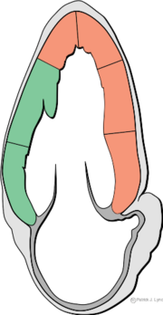 |
15 KB | 1 | |
| 15:21, 3 October 2007 | A5Cnormal.avi (file) | 465 KB | A 5 chamber view | 1 | |
| 15:09, 1 October 2007 | A2Cnormal.avi (file) | 392 KB | Apical 2 chamber view of a normal heart | 1 | |
| 15:03, 1 October 2007 | A3Cnormal.avi (file) | 499 KB | Apical 3 chamber view of a normal heart | 1 | |
| 14:59, 1 October 2007 | PLAXnormal.avi (file) | 214 KB | Parasternal long axis view of a normal heart | 1 | |
| 14:55, 1 October 2007 | SAXAP normal.avi (file) | 314 KB | A parasternal short axis on apical level | 1 | |
| 14:42, 1 October 2007 | SAXPAP normal.avi (file) | 371 KB | A parasternal short axis view on level of the papillary muscle | 1 | |
| 14:40, 1 October 2007 | Subcostal normal.avi (file) | 159 KB | Subcostal view of a normal heart | 1 | |
| 14:35, 1 October 2007 | SuprasternalNormal.avi (file) | 254 KB | Suprasternal view of a normal heart | 1 | |
| 11:24, 1 October 2007 | A4C normal.avi (file) | 207 KB | Apical 4 Chamber view of a normal heart | 1 | |
| 13:47, 25 September 2007 | PLAXLynch.png (file) | 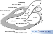 |
64 KB | Parasternal long axis view | 1 |
| 13:35, 25 September 2007 | Torso.png (file) | 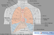 |
87 KB | Torso Schematic | 1 |
| 13:03, 25 September 2007 | EdlerHertz.png (file) | 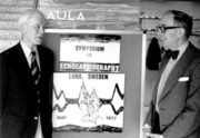 |
39 KB | Inge Edler and Hellmuth Hertz in 1977 in the University Hospital in Lund. | 1 |
| 12:57, 25 September 2007 | FirstEchoCor.png (file) | 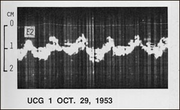 |
54 KB | The first echocardiogram made by Inge Edler and Hellmuth Hertz in 1953 | 1 |
| 13:13, 24 September 2007 | A4CTTS.avi (file) | 619 KB | Apical 4 chamber view of a Takotsubo cardiomyopathy | 1 | |
| 12:13, 24 September 2007 | MVThrombus.avi (file) | 709 KB | 1 | ||
| 11:17, 24 September 2007 | PulsusAlternans.png (file) |  |
1.27 MB | An ECG of a 40 year old man with Kahlers' disease and tamponade. Note the electrical alternans in the precordial leads and the microvoltages in the extremity leads | 1 |
| 09:44, 24 September 2007 | Swingingheart.avi (file) | 1 MB | Apical 4 Cjaner view of a swinging heart. Note the pericardial effusion. | 1 |