| HOCM with SAM
|
|
|
| Courtesy of: AMC Echolab, AMC, The Netherlands
|
| <html5media height="350" width="279" autoplay="true" loop="true">File:E00502.mp4</html5media>
|
<html5media height="350" width="279" autoplay="true" loop="true">File:E00503.mp4</html5media>
|
| PLAX: concentric hypertrophy of left ventricle, SAM of AMVL
|
PLAX with Color Doppler: high velocity turbulent flow in LVOT
|
 enlarge enlarge
 download file to use in your powerpoint presentation download file to use in your powerpoint presentation
|
 enlarge enlarge
 download file to use in your powerpoint presentation download file to use in your powerpoint presentation
|
|
|
| <html5media height="350" width="279" autoplay="true" loop="true">File:E00504.mp4</html5media>
|
<html5media height="350" width="279" autoplay="true" loop="true">File:E00505.mp4</html5media>
|
| A4CH: concentric hypertrophy of left ventricle, SAM of AMVL
|
A3CH
|
 enlarge enlarge
 download file to use in your powerpoint presentation download file to use in your powerpoint presentation
|
 enlarge enlarge
 download file to use in your powerpoint presentation download file to use in your powerpoint presentation
|
|
|
| <html5media height="350" width="279" autoplay="true" loop="true">File:E00506.mp4</html5media>
|
<html5media height="350" width="279" autoplay="true" loop="true">File:E00507.mp4</html5media>
|
| A3CH with Color Doppler: high velocity turbulent flow in LVOT
|
A4CH with Color Doppler: high velocity turbulent flow in LVOT and in the middle of left ventricle
|
 enlarge enlarge
 download file to use in your powerpoint presentation download file to use in your powerpoint presentation
|
 enlarge enlarge
 download file to use in your powerpoint presentation download file to use in your powerpoint presentation
|
|
|
|
|
|
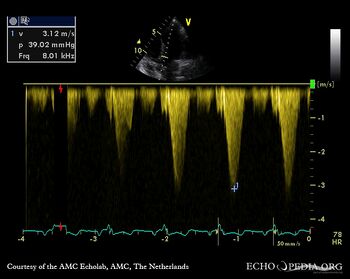
| Continuous-wave doppler signal: dynamic gradient in LVOT
|
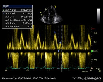
Pulsed-wave doppler signal of transmitral flow
|
|
|
|
|
|
|
|
|
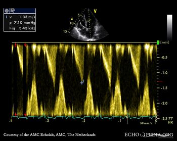
| Continuous-wave doppler signal: dynamic gradient in the middle of left ventricle
|
|
|
|
|


